
Embark on a scientific voyage with our comprehensive guide, “Color the Bone Matrix Answer Key,” where we unravel the mysteries of bone matrix coloration, a crucial technique in histological analysis. This guide delves into the significance of staining techniques, empowering you to decipher the composition and mineralization of bone tissue with unparalleled precision.
Our journey begins with an exploration of the diverse staining methods employed to visualize bone matrix, providing a solid foundation for understanding their applications. We then embark on a detailed examination of commonly used stains, deciphering their specificities and target tissues.
Introduction to Bone Matrix Coloration
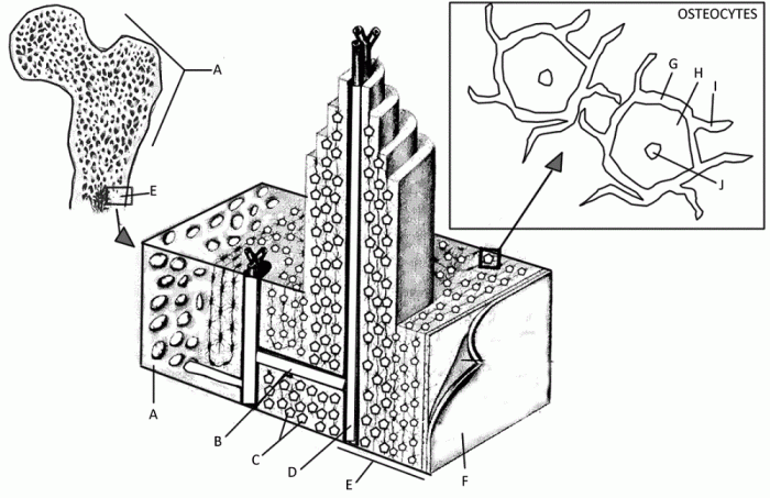
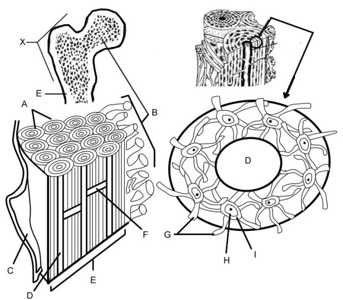
Bone matrix coloration is a crucial technique in histological analysis, providing valuable insights into the structure and composition of bone tissue. By employing specific staining methods, researchers can differentiate between various components of the bone matrix, such as collagen fibers, mineralized areas, and osteocytes.
A range of staining techniques are available for bone matrix coloration, each with its unique advantages and applications. These techniques utilize dyes or stains that selectively bind to specific components of the bone matrix, allowing for their visualization under a microscope.
Staining Techniques for Bone Matrix Coloration
- Hematoxylin and Eosin (H&E) Staining:A basic staining method that differentiates between nuclei (stained blue) and cytoplasm (stained pink), providing a general overview of bone tissue.
- von Kossa Staining:A silver-based staining technique that stains mineralized areas of the bone matrix black, highlighting the distribution of calcium salts.
- Masson’s Trichrome Staining:A versatile staining method that differentiates between collagen fibers (stained blue), muscle fibers (stained red), and nuclei (stained black), providing detailed information about the bone matrix architecture.
Types of Bone Matrix Stains
Bone matrix stains are histological stains used to visualize and differentiate various components of the bone matrix, such as collagen, calcium, and other minerals. These stains are commonly employed in bone research and diagnostics to assess bone structure, mineralization, and pathology.
The choice of stain depends on the specific target tissue or matrix component of interest. Some commonly used bone matrix stains include hematoxylin, eosin, Masson’s trichrome, and alizarin red.
Bone Matrix Stain Types
| Stain Name | Color | Target Tissue | Applications |
|---|---|---|---|
| Hematoxylin | Blue | Nuclei | Nuclear staining in bone cells |
| Eosin | Pink | Cytoplasm | Cytoplasmic staining in bone cells |
| Masson’s Trichrome | Blue, Red, Yellow | Collagen, Muscle, Keratin | Differentiating between collagen fibers and other matrix components |
| Alizarin Red | Orange-Red | Calcium Deposits | Mineralization assessment in bone tissue |
Procedures for Bone Matrix Staining: Color The Bone Matrix Answer Key
Bone matrix staining is a crucial technique in histology for visualizing and studying the structure and composition of bone tissue. The general steps involved in bone matrix staining include tissue preparation, staining reagents, incubation times, and rinsing procedures.
Tissue preparation involves decalcification, which removes the mineral components of the bone, leaving behind the organic matrix. This step is essential for allowing the staining reagents to penetrate the tissue and bind to the specific components of interest.
Staining Reagents
A variety of staining reagents are used for bone matrix staining, each targeting specific components. Some common reagents include:
- Hematoxylin and eosin (H&E) stain: Provides a general overview of the tissue structure, highlighting nuclei (blue) and cytoplasm/matrix (pink).
- Von Kossa stain: Stains calcium deposits black, making it useful for visualizing mineralized bone matrix.
- Alizarin red S stain: Binds to calcium and stains mineralized bone matrix red.
- Goldner’s trichrome stain: Differentiates between collagen fibers (blue) and osteoid (red), which is unmineralized bone matrix.
Incubation Times
Incubation times vary depending on the staining reagent used. Generally, tissues are incubated in the staining solution for a specific duration to allow the reagent to bind to the target components. Incubation times can range from a few minutes to several hours.
Rinsing Procedures
After staining, tissues are rinsed thoroughly to remove excess staining solution and prevent background staining. Rinsing is typically done using distilled water or a buffer solution.
Interpretation of Bone Matrix Coloration

Bone matrix coloration can provide valuable information about the composition and mineralization of the bone tissue. The color of the bone matrix can vary depending on the type of stain used and the presence of pathological conditions.
When bone is stained with hematoxylin and eosin (H&E), the mineralized bone matrix typically appears pink or red. This is because the calcium salts in the bone matrix bind to the eosin stain, which is a negatively charged dye. The unmineralized bone matrix, such as osteoid, appears blue or purple because it binds to the hematoxylin stain, which is a positively charged dye.
Use of Bone Matrix Coloration to Identify Pathological Conditions
Bone matrix coloration can also be used to identify pathological conditions. For example, in osteomalacia, a condition in which the bone matrix is not properly mineralized, the bone matrix may appear blue or purple on H&E staining. This is because the unmineralized bone matrix binds more strongly to the hematoxylin stain than to the eosin stain.
Troubleshooting Common Issues
Bone matrix staining is a technique that can be challenging to master. Several common problems can occur during the staining process, leading to unsatisfactory results. Troubleshooting these issues is essential for obtaining accurate and reliable staining results.
Incorrect Staining Times, Color the bone matrix answer key
- Problem:Overstaining or understaining of the bone matrix.
- Troubleshooting:Adjust the staining times according to the specific staining protocol and the thickness of the tissue section. Use a microscope to monitor the staining process and stop the reaction when the desired intensity is achieved.
Insufficient Rinsing
- Problem:Background staining or uneven staining of the bone matrix.
- Troubleshooting:Ensure thorough rinsing of the tissue sections between each step of the staining procedure. Use copious amounts of distilled water or buffer to remove excess stain and prevent the accumulation of salts or other contaminants.
Tissue Damage
- Problem:Loss of tissue integrity or disruption of the bone matrix.
- Troubleshooting:Handle the tissue sections gently throughout the staining process. Use appropriate fixatives and decalcifying agents to preserve the tissue structure. Avoid excessive agitation or prolonged exposure to harsh chemicals.
Non-Specific Staining
- Problem:Staining of non-target structures or background staining.
- Troubleshooting:Optimize the staining protocol by adjusting the pH, ionic strength, and concentration of the staining solutions. Use blocking agents or counterstains to minimize non-specific interactions.
Clarifying Questions
What is the significance of bone matrix coloration in histological analysis?
Bone matrix coloration enables researchers to visualize and differentiate various bone components, such as collagen fibers and mineralized areas, providing valuable insights into bone structure and composition.
How does the color of the bone matrix provide information about its composition and mineralization?
The color imparted by different stains corresponds to specific bone matrix components. For instance, hematoxylin stains nuclei blue, while alizarin red stains calcium deposits red, allowing researchers to assess bone mineralization and composition.
What are some common issues encountered in bone matrix staining and how can they be resolved?
Common issues include incorrect staining times, insufficient rinsing, and tissue damage. To troubleshoot, adjust staining durations, ensure thorough rinsing, and handle tissues gently during the staining process.
